


| Protocol PDF | Click for Protocol |
|---|---|
| Catalog Number | TK01343 |
| Species | Human |
| Design | Indirect solid phase enzyme immunometric assay (ELISA). |
| Standards | One cut-off control, ready to use. |
| Controls | Negative control, positive control, and strong positive control, ready to use. |
| Sample Types | Human serum or plasma. |
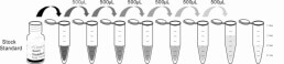
| Sample Volume | 100 μL of properly diluted (1:100) unknown / determination. |
| Assay Desc. | 30 min. incubation (RT) + 15 min. (RT) + 15 min. (RT) + 5 min. (RT) = 1 hour, 5 min. total incubation time. |
| Standard Range | Evaluation is carried out by direct comparison of the optical density of each sample with the optical density of the controls. |
| Sensitivity | |
| Radioactivity | |
| Other Names | |
| Storage | 2 - 8°C |
| Postscript | For research use only, not for use in diagnostic procedures. |
This ELISA kit applies to the in vitro quantitative determination of Anti-β2 Glycoprotein I Screen concentrations in serum, plasma and other biological fluids.
This ELISA kit uses the Sandwich-ELISA principle. The micro ELISA plate provided in this kit has been pre-coated with an antigen specific to Anti-β2 Glycoprotein I Screen. Standards or samples are added to the micro ELISA plate wells and combined with the specific antigen. Then a biotinylated detection antigen specific for Anti-β2 Glycoprotein I Screen and Avidin-Horseradish Peroxidase (HRP) conjugate are added successively to each micro plate well and incubated. Free components are washed away. The substrate solution is added to each well. Only those wells that contain Anti-β2 Glycoprotein I Screen, biotinylated detection antigen and Avidin-HRP conjugate will appear blue in color. The enzyme-substrate reaction is terminated by the addition of stop solution and the color turns yellow. The optical density (OD) is measured spectrophotometrically at a wavelength of 450 nm ± 2 nm. The OD value is proportional to the concentration of Anti-β2 Glycoprotein I Screen. You can calculate the concentration of Anti-β2 Glycoprotein I Screen in the samples by comparing the OD of the samples to the standard curve.
As the OD values of the standard curve may vary according to the conditions of the actual assay performance (e.g. operator, pipetting technique, washing technique or temperature effects), the operator should establish a standard curve for each test. Typical standard curve and data is provided below for reference only.
Intra-assay Precision (Precision within an assay): 3 samples with low, middle,high concentration of Anti-β2 Glycoprotein I Screen. And the sample of Anti-β2 Glycoprotein I Screen were tested 30 times on one plate, respectively. Inter-assay Precision (Precision between assays): 3 samples with low, middle,high concentration of Anti-β2 Glycoprotein I Screen. And the sample of Anti-β2 Glycoprotein I Screen were tested on 3 different plates, 30 replicates in each plate.
Matrices listed below were spiked with certain level of Anti-β2 Glycoprotein I Screen and the recovery rates were calculated by comparing the measured value to the expected amount of Anti-β2 Glycoprotein I Screen in samples.
| Matrix | Recovery range (%) | Average Recovery (%) |
|---|---|---|
| Serum (n=5) | 91-107 | 96 |
| EDTA plasma (n=5) | 91-98 | 94 |
| heparin plasma (n=5) | 84-97 | 93 |
The linearity of the kit was assayed by testing samples spiked with appropriate concentration of Anti-β2 Glycoprotein I Screen and their serial dilutions. The results were demonstrated by the percentage of calculated concentration to the expected.
| Serum (n=5) | EDTA plasma (n=5) | heparin plasma (n=5) | ||
|---|---|---|---|---|
| 1:2 | Range (%) | 89-101 | 91-101 | 91-107 |
| Average (%) | 93 | 93 | 95 | |
| 1:4 | Range (%) | 91-107 | 92-105 | 94-108 |
| Average (%) | 95 | 95 | 94 | |
| 1:8 | Range (%) | 93-101 | 88-108 | 93-108 |
| Average (%) | 93 | 95 | 93 | |
| 1:16 | Range (%) | 93-108 | 89-108 | 89-104 |
| Average (%) | 94 | 94 | 93 |
The stability of Anti-β2 Glycoprotein I Screen is determined by the loss rate of activity. The loss rate of Anti-β2 Glycoprotein I Screen is less than 5% within the expiration date under appropriate storage condition. To minimize extra influence on the performance, operation procedures and lab conditions, especially room temperature, air humidity, incubator temperature should be strictly controlled. It is also strongly suggested that the whole assay is performed by the same operator from the beginning to the end.
| Item | Specifications | Storage |
|---|---|---|
| Micro ELISA Plate(Dismountable) | 8 wells ×12 strips | -20℃, 6 months |
| Reference Standard | 2 vials | |
| Concentrated Biotinylated Detection Ab (100×) | 1 vial, 120 μL | |
| Concentrated HRP Conjugate (100×) | 1 vial, 120 μL | -20℃(shading light), 6 months |
| Reference Standard & Sample Diluent | 1 vial, 20 mL | 4℃, 6 months |
| Biotinylated Detection Ab Diluent | 1 vial, 14 mL | |
| HRP Conjugate Diluent | 1 vial, 14 mL | |
| Concentrated Wash Buffer (25×) | 1 vial, 30 mL | |
| Substrate Reagent | 1 vial, 10 mL | 4℃(shading light) |
| Stop Solution | 1 vial, 10 mL | 4℃ |
| Plate Sealer | 5 pieces | |
| Product Description | 1 copy | |
| Certificate of Analysis | 1 copy |
Microplate reader with 450 nm wavelength filter
High-precision transfer pipette, EP tubes and disposable pipette tips
Incubator capable of maintaining 37℃
Deionized or distilled water
Absorbent paper
Loading slot for Wash Buffer
1. Prepare all reagents, samples and standards;
2. Add 100µL standard or sample to each well. Incubate 1 hours at 37°C;
3. Aspirate and add 100µL prepared Detection Reagent A. Incubate 1 hour at 37°C;
4. Aspirate and wash 3 times;
5. Add 100µL prepared Detection Reagent B. Incubate 30 minutes at 37°C;
6. Aspirate and wash 5 times;
7. Add 90µL Substrate Solution. Incubate 10-20 minutes at 37°C;
8. Add 50µL Stop Solution. Read at 450nm immediately.

Featured Products
- STK3, 1-322aa, Human, His-tag, Baculovirus
- Human MIP-5(Macrophage Inflammatory Protein 5) ELISA Kit
- Human MIF(Macrophage Migration Inhibitory Factor) ELISA Kit
- Human FZD8(Frizzled Homolog 8) ELISA Kit
- Human PDCD1(Programmed Cell Death Protein 1) ELISA Kit
- Human CRHBP(Corticotropin Releasing Hormone Bingding Protein) ELISA Kit
- Human αHSP(Alpha-Hemoglobin Stabilizing Protein) ELISA Kit
- Human MMP-28(Matrix Metalloproteinase 28) ELISA Kit
- Human TLR4(Toll-Like Receptor 4) ELISA Kit
- Human SPTAN1(Alpha-Fodrin) ELISA Kit
- Human AZGP1(Zinc-alpha-2-glycoprotein) ELISA Kit
- Human MSLN(Mesothelin) ELISA Kit
- Human SOST(Sclerostin) ELISA Kit
- Mab21 Domain Containing Protein 1 (MB21D1)
- Coiled Coil Domain Containing Protein 60 (CCDC60)
© Copyright 2024 TZYBIOTECH. All Rights Reserved. SiteMap